(CS-141) Point-of-Care Multimodal Imaging in Mobile Chronic Wound Care: Insights from a Case Series
Friday, April 10, 2026
Introduction:
Chronic wounds represent a silent epidemic, affecting approximately 10.5 million Medicare beneficiaries in the United States and imposing an annual cost of $22.5 billion on Medicare alone.1 In mobile wound care settings, limited access to advanced diagnostics often delays the detection of perfusion deficits, infections, and atypical pathologies.2 Multimodal imaging — integrating digital photography, near-infrared spectroscopy (NIRS), and thermography — offers an innovative point-of-care solution, providing real-time assessment of tissue oxygenation (StO₂) and temperature as a surrogate marker for infection. This case series investigates the clinical utility of a multimodal imaging in guiding interventions to optimize outcomes for chronic wounds in mobile care environments.
Methods: Six adult patients with chronic wounds, including venous stasis ulcers, post-amputation stumps, atypical calf wounds, and lymphedema-associated wounds, were evaluated in mobile care settings. Patients had multiple comorbidities, such as peripheral vascular disease, arthritis, cellulitis, microvascular disease, and edema. A pocket-sized multimodal imaging device* was used at the point of care to assess wound characteristics.
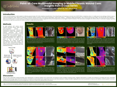
Results: Multimodal imaging provided actionable insights across all six patients by differentiating tissue perfusion, infection, and atypical wound conditions. In wounds with adequate perfusion, such as the venous stasis ulcer, mean wound bed StO₂ exceeded 50% with heterogeneous periwound oxygenation, indicating strong healing potential. Cases exhibiting early compromise demonstrated elevated periwound temperature and oxygenation (97–100%), allowing proactive management before overt tissue injury. Imaging also helped rule out vascular deficits and redirect diagnoses in atypical wounds, as seen in the posterior calf case diagnosed with mycosis fungoides. For wounds with evolving perfusion deficits, serial assessments enabled timely interventions to mitigate complications. Poorly perfused wounds showed limited healing potential, prompting vascular referral and close monitoring. Thermography aided in detecting infection and monitoring treatment response, with localized hotspots (ΔT >4°F) observed even after infection resolution.
Discussion: Point-of-care multimodal imaging facilitates early identification of perfusion deficits, skin compromise, and atypical wound conditions, allowing timely, targeted interventions in mobile care settings. By providing real-time, actionable insights, this technology helps overcome limitations of traditional visual assessments, supporting more accurate clinical decision-making. Portable imaging devices have the potential to improve care delivery, enhance patient outcomes, and promote equitable, efficient wound management in resource-limited and underserved environments.

.jpg)