(CR-045) A comprehensive Scoping Review on the Use of Point-of-care Infrared Thermography Devices for Assessing Diabetic Foot Ulcers
Friday, April 10, 2026
Heba Tallah Mohammed, MD,PhD – Swift Medical Inc.; Amy Cassata, BSc; Robert D. J. Fraser, BScN,MN – Swift Medical Inc.
Introduction: This scoping review synthesized evidence from thirteen studies on point-of-care infrared thermography devices for diabetic foot ulcers (DFUs), including prospective cohorts, cross-sectional comparisons, and pilot series (Level III–IV evidence). We descriptively summarized findings on diagnostic thresholds, which showed thermography’s role in predicting healing, identifying complications, and monitoring DFUs.
Methods: A scoping review was conducted following Arksey & O’Malley and PRISMA-ScR guidelines. Medline, Embase, CINAHL, and Cochrane Library were searched for human studies evaluating point-of-care infrared thermography devices in DFU wound assessment, with all study designs included. Data was extracted from 13 eligible studies, capturing study design, sample size, device type, outcomes, and key findings. Findings were synthesized descriptively and presented in narrative and tabular formats.
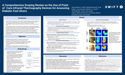
Results: The 13 DFU studies (median n=24, range 1-100) were predominantly small observational cohorts or case series (Level III–IV evidence), with 7 out of 13 (54%) studies including a control group. Thermography detected early thermal changes in acute DFUs before they were clinically visible or symptomatic, and before complications were evident.1 Thermography also demonstrated potential in detecting success of post-revascularization treatment.2 In many studies, thermography was shown to identify chronic temperature increases associated with infection risk in DFUs.3,4 Additionally, monitoring weekly thermographic changes accurately predicted healing trajectory (p=0.036).5 Furthermore, 3D thermography detected wound inflammation and helped predict DFU ulceration risk.6 Challenges were identified in integrating thermography into clinical practice. These included reimbursement, training needs, EMR integration, and the absence of standardized protocols (particularly for skin tone variability). However, despite these limitations, studies broadly supported thermography’s role in early complication detection and real-time monitoring.
Discussion: Thermography is a convenient, non-invasive tool for DFU management that can improve monitoring and enable early detection of complications. Although current evidence is largely Level III-IV, advancements in EMR integration, standardized protocols, and clinician training are still needed to establish thermography as a standard clinical tool in DFU care.

.jpg)