
.jpg)
Laboratory Research
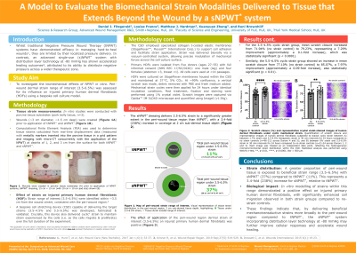
Wounded (~3 cm diameter, ~1.5 cm depth) porcine tissue models (n=3) were treated with sNPWT* and tNPWT† (with foam filler). Tissue strain was quantified using Finite Element Analysis (FEA) based on displacement data from micro-CT imaging of metallic markers embedded at various depths. Strain range of interest was identified within the peri-wound region. A bespoke cell stretching device (CSD) was developed to deliver the strain to cultured HDFs on collagen I-coated elastic membranes. HDFs from five donors with full informed consent (NHS REC 17/SC/0220) were cultured to confluence, injured via scratch assay, and then subjected to strain (3.5–4.5% and 5.5–6.5%) using the CSD for 24 h. Post-treatment, scratch closure was quantified using crystal violet staining and image analysis. Statistical analysis was performed using Welch’s t-test.
Results:
The peri-wound tissue region experiences a 3.4-fold (238%) increase in coverage in the strain range of interest (3.5-6.5%) with sNPWT* compared to tNPWT† at a sub-dermal depth of 2 cm. Strain application enhanced HDF migration: 3.5–4.5% strain improved scratch closure by 7.29% (p< 0.0001), and 5.5–6.5% strain by 7.97% (p< 0.01), compared to no-strain controls.
Discussion:
The strain ranges of interest were associated with enhanced cell migration in vitro. These findings indicate that, by delivering beneficial mechanotransductive strains more broadly to the peri-wound region compared to tNPWT†, the sNPWT* system incorporating distribution layer technology at -80 mmHg may further improve cellular responses and accelerate wound healing.