
.jpg)
Case Series/Study
Lower-extremity diabetic ulcers (LEDUs) are a frequent and highly morbid complication of diabetes,
with infection representing a major driver of hospitalization in low-resource settings. This case report
describes the healing response of a hard-to-heal LEDU in an elderly female with poorly controlled
diabetes (A1c >10%) following wound bed preparation using a topical dehydrating agent (TDA*) and
subsequent treatment with a wool-derived keratin-based matrix (KBM†) xenograft.
Methods:
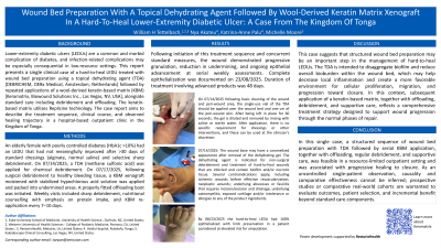
The case was managed in a hospital-based outpatient wound clinic in the Kingdom of Tonga. Prior to
advanced intervention, the ulcer was treated for >30 days with standard dressings (alginate, normal
saline) and selective sharp debridement without meaningful improvement. On 14/07/2025, a TDA,
containing methane sulfonic acid, was applied for chemical debridement. On 17/07/2025, following
surgical debridement to healthy bleeding tissue, a KBM xenograft moistened with stabilized
hypochlorous acid solution was applied and packed into undermined areas. A properly fitted
offloading boot was also initiated. Weekly clinic visits included sharp debridement, nutritional
counseling with emphasis on protein intake, and re-application of the KBM every 7–10 days.
Results:
After initiation of TDA and routine KBM applications, the wound demonstrated progressive
granulation, reduction in undermining, and sustained epithelial advancement. Complete
epithelialization was achieved by 23/08/2025. Total length of treatment with advanced
products, 48 days.
Discussion:
This case illustrates that combining a TDA with routine application of a KBM xenograft can
jump-start wound progression toward closure in a hard-to-heal LEDU. This approach may
represent a valuable strategy in resource-limited environments where timely wound bed
preparation and biologic support are essential for achieving optimal outcomes.