
.jpg)
Clinical Research
Visual inspection remains the primary approach for assessing wound tissue types such as granulation, slough, eschar, and epithelialization. As clinicians rely on their own experience, training, and situational judgment, visual interpretation can vary, resulting in inconsistent assessments.1 Artificial intelligence (AI)–based segmentation may help reduce this variability by standardizing visual representations.2 This study explored how clinicians identify and estimate tissue types when reviewing AI-captured images and compared their assessments with AI-generated measurements to understand where differences persist.
Methods:
A cross-sectional survey of 50 clinicians was done through conference outreach and professional networks. Participants answered questions on their demographics and experience in wound care, then nine anonymized images of wounds with a wide range of tissue, types, and skin tones were shown to clinicians. For each image, clinicians indicated the presence of each tissue type and offered their best estimate of the proportion of each. These estimates were then compared with values generated by the AI-based segmentation tool, and agreement was examined using intraclass correlation coefficients (ICCs). Pearson correlations were used to assess alignment between clinicians and AI.
Results:
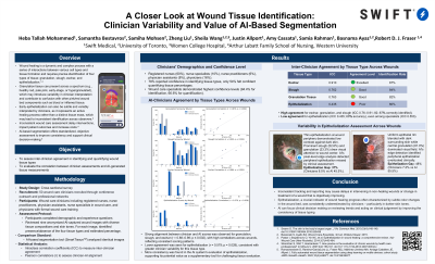
The sample included registered nurses (60%), nurse specialists (10%), nurse practitioners (6%), physician assistants (8%), and physicians (16%). Wound care specialists had the highest confidence in ability to identify (94.4%) and quantify (66.6%) different tissue types.
There was greatest agreement among clinicians for granulation, slough, and eschar, viewing them in 82%, 84%, and 87% of images, respectively, and their values closely agreed with AI values (r=0.879–0.984; ICC 0.76–0.91).
On the other hand, the percentage of correct interpretation of epithelialization was relatively low, with only 60% of the images being correctly identified by clinicians, and a low ICC of 0.39- 0.55. Even for specialists in wound care, agreement was rather low, with an ICC of 0.553. Correlation with AI was moderate and AI generated more consistent estimates.
Discussion:
Epithelization was the most difficult area to assess. With its thinning nature that integrates well with surrounding skin, it has been easy for clinicians to underestimate. The use of AI can support clinicians in providing more consistent assessment of this type.