
.jpg)
Laboratory Research
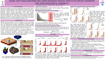
A multilayer sacral finite element model was developed in Abaqus/CAE 2024 that included epidermis, dermis, adipose tissue, and muscle with literature-based thicknesses and mechanical properties [1,2]. Tissue stiffness was varied between softer tissues, associated with aging or degeneration, and stiffer tissue states. Two device geometries were simulated: a round, flat sensor and a narrow, elongated cable. Uniform pressures of 2-10 kPa were applied beneath each device. Stress and strain distributions were evaluated within a 30-mm region of interest, and the top quartile of strain values was used to compare tissue exposure across device types and tissue conditions.
Results:
The two device geometries produced distinct tissue deformation patterns. Under 10 kPa loading, the flat, round sensor generated relatively uniform strain fields, while the narrow cable created localized regions of concentrated strain. This resulted in 1.25-fold more tissue being in the highest strain quartile for the cable compared with the sensor. Softened tissues increased tissues exposed to high strain 1.4-fold for the sensor and 1.6-fold for the cable. Stiffened tissues decreased the amount of tissue in the highest strain quartile by 1.1-fold for the sensor and 1.3-fold for the cable.
Discussion:
Device geometry and tissue stiffness together influence mechanical loading effects in sacral soft tissues. The cable exposed more tissue to high strains than the flat sensor, and this effect increased with tissue softening. Stiffened tissues reduced tissue exposed to high strain for both device types. This highlights the importance of considering tissue variability and device geometry when assessing medical device related pressure injury risk and in design of wearable technologies.