(CR-043) Correlation of Multispectral Near-Infrared Imaging with Standard Vascular Diagnostics in Chronic Wound Care
Friday, April 10, 2026
Adam Iddriss, MD; Star-Kayla Lewis, MD; Natasjia Pinnock, MD; Marisa Ranire-Maguire, MD; Amit Rao, MD
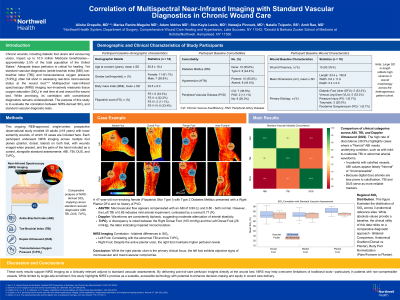
Introduction: Chronic wounds including diabetic foot ulcers and venous leg ulcers impact up to 10.5 million of Medicare beneficiaries 2.5% of the total population of the United States.1 Adequate tissue perfusion is critical for healing.2,3 Yet standard vascular diagnostics (ankle-brachial index (ABI), toe-brachial index (TBI), and transcutaneous oxygen pressure (TcPO₂)) often fall short in assessing real-time, microvascular status at the wound bed.4 Multispectral near-infrared spectroscopy (NIRS) imaging non-invasively measures tissue oxygen saturation (StO₂) in real time at and around the wound bed. While promising, its correlation with conventional diagnostics remains underexplored. The purpose of this study is to evaluate the correlation between NIRS-derived StO₂ and standard vascular diagnostic tests.
Methods: This ongoing REB-approved, single-center, prospective observational study enrolled 40 adults (≥18 years) with lower extremity wounds, of which 11 cases are included here. Each participant underwent NIRS imaging* across multiple foot planes (plantar, dorsal, lateral) on both feet, with wounds imaged when present, and the palm of the hand included as a control, alongside standard assessments: ABI, TBI, and TcPO₂. Primary analysis used Spearman correlation to assess StO₂ relationships with vascular parameters (p< 0.05). Secondary endpoints included stratification by PAD (Rutherford, Fontaine, WIfI), venous disease (CEAP), and wound severity (Texas classification). Exploratory analyses examined StO₂ variability by age, sex, diabetes status, and Fitzpatrick skin type.
Results: The data demonstrated a strong correlation between NIRS-derived StO₂ and TcPO₂, with TCOM < 40 mmHg corresponding to StO₂ < 39% range. Moderate correlations were observed with ABI; however, in patients with diabetes—where ABI is known to be unreliable5 — StO₂ showed stronger correlation with TBI. Importantly, NIRS achieved high imaging success rates, likely due to its non-contact and non-invasive design.
Discussion: These early results support NIRS imaging as a clinically relevant adjunct to standard vascular assessments. By delivering point-of-care perfusion insights directly at the wound bed, NIRS may help overcome limitations of traditional tools—particularly in patients with non-compressible vessels. While limited by single-site enrollment, this study highlights NIRS’s promise as a scalable, accessible technology with potential to enhance decision-making and equity in wound care delivery.

.jpg)