
.jpg)
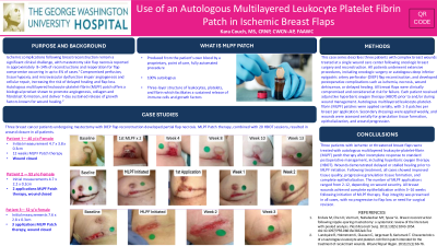
Case Series/Study
Ischemic complications following breast reconstruction remain a significant clinical challenge, with mastectomy skin flap necrosis reported in approximately 8–14% of reconstructions and reoperation for flap compromise occurring in up to 4% of cases. ¹ Compromised perfusion, tissue hypoxia, and microvascular dysfunction impair angiogenesis and cellular repair, increasing the risk of delayed healing and flap loss. Autologous multilayered leukocyte-platelet-fibrin (MLPF) patch offers a biological product shown to promote angiogenesis, collagen and fibroblast formation, and deliver 7-day sustained release of growth factors known for wound healing.2
Methods:
This case series describes three patients with complex breast wounds treated at a single wound care center following oncologic breast surgery and reconstruction. All patients underwent extensive procedures, including oncologic surgery or autologous deep inferior epigastric artery perforator (DIEP) flap reconstruction, and developed postoperative complications such as ischemia, necrosis, wound dehiscence, or delayed healing. All breast flaps were clinically compromised and considered at risk for failure. Each patient received adjunctive hyperbaric oxygen therapy (HBOT) prior to and/or during wound management. Autologous multilayered leukocyte-platelet-fibrin (MLPF) patches were applied serially, with 1-3 patches per breast per application. Secondary dressings were applied weekly, and wounds were assessed serially for granulation tissue formation, epithelialization, and wound progression.
Results:
Three patients with ischemic or threatened breast flaps were treated with autologous multilayered leukocyte-platelet-fibrin (MLPF) patch therapy after incomplete response to standard postoperative management, including hyperbaric oxygen therapy (HBOT). Wounds demonstrated delayed or stalled healing prior to MLPF initiation. Following treatment, all cases showed improved tissue quality, progressive granulation tissue formation, and complete epithelialization. The number of MLPF applications ranged from 2-12, depending on wound severity. All breast wounds achieved complete epithelialization within 3–16 weeks following initiation of MLPF therapy. Flap integrity was preserved in all cases, with no progression to flap loss or need for surgical revision.
Discussion:
In this case series, autologous multilayered leukocyte-platelet-fibrin (MLPF) patch therapy was associated with successful healing of ischemic breast flaps refractory to standard postoperative management, including hyperbaric oxygen therapy. By providing an angiogenic, native cellular scaffold within ischemic tissue, MLPF may help mitigate flap failure risk and support durable wound closure in complex reconstructive cases.