
.jpg)
Case Series/Study
Limb salvage in the setting of osteomyelitis often requires osseous resection, yet the resulting structural voids and compromised soft-tissue environment can predispose patients to recurrent infection, delayed healing, and progressive deformity or shortening of the foot. Biological grafts that mimic native extracellular matrix may help stabilize the post-resection space, support tissue regeneration, and maintain foot architecture. Intact fish skin grafts*, rich in omega-3 fatty acids, represent a promising biomaterial for promoting healing in contaminated or previously infected fields. This study evaluates the utility of placing fenestrated intact fish skin graft* into osseous defects immediately after infected bone removal in limb-salvage procedures.
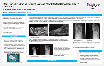
Methods: Patients who were identified with osteomyelitis that were going to undergo primary bone resection and closure over that wound were enrolled longitudinally. All patients were treated in the operating room with regional or general anesthesia. All patients had perioperative antibiotics tailored to the residual bone margin pathology. All patients underwent a single stage surgical incision and removal of the infected bone(s). Fenestrated intact fish skin* graft was then rolled and placed into the defect left by the excised bone. The primary incision was closed with vertical mattress nonabsorbable suture. Negative pressure was not employed.
Results: We present postoperative post-operative radiographs and MRIs of 3 of 13 patients treated in this manner. All 13 patients went on to complete healing over the course of 8 weeks. Interestingly digit and foot length was maintained with this technique. No patient required repeat bone resection or additional surgical intervention for infection control. These outcomes suggest that adjunctive acellular fish skin graft* placement may help stabilize the post-resection environment, supporting soft-tissue regeneration and preventing infection. In some radiographs what appears to be bony replacement of the margins of the fish skin can be seen.
Discussion: The preserved extracellular matrix structure and omega-3–rich lipid content of the fish-skin graft may contribute to angiogenesis, modulation of local inflammation, and enhanced cellular ingrowth, which are mechanisms that may be especially beneficial following osseous resection in contaminated or compromised fields. The fact that digital and foot length is maintained even at 16 weeks post-resection is notable.