(PI-036) A portable photoacoustic platform for real-time deep tissue oxygenation.
Friday, April 10, 2026
Carolyn Bayer, Ph.D.; Miller Dickerson, N/A; Mistina Mano Manoharan, MBBS; Brandon Moore, N/A
Introduction: Peripheral tissue oxygenation is a critical indicator of wound health and healing. Standard wound care approaches rely solely on indirect assessment of flow and visual assessments of wound morphology. Oximetry based technologies such as transcutaneous oximetry, near-infrared spectroscopy, or spatial frequency domain imaging are bulky, expensive, and require long calibration times to assess tissue oxygenation at shallow imaging depths (~1-2 mm). This makes tissue oxygenation assessments inaccessible as part of routine wound care in traditional high volume low-resources clinics. Spectral photoacoustic imaging (sPAI) is a rapidly emerging hybrid modality that utilizes pulsed light to generate acoustic waves detectable with conventional ultrasound hardware. The use of pulse light with unique wavelengths allows direct mapping of light absorbing chromophores such as oxy- & deoxy-hemoglobin.
Methods: Recently, our team at LumaWave, developed a portable sPAI system for real-time tissue oximetry capable of imaging up to depths of ~4 cm in vivo. In this study, we present a case study where this prototype was demonstrated in humans to measure microvascular & arterial oxygenation in the forearm.
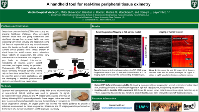
Results: We demonstrated hemoglobin specific imaging up to a depth of 3.5 cm with clear visualization of the radial artery. Additionally, we demonstrated oxygenation mapping in real-time.
Discussion:
Our results demonstrate the ability of our platform technology to provide real-time peripheral tissue oximetry that allows monitoring of wound healing, revolutionizing wound care and peripheral tissue health.

.jpg)